|
Seborrheic
dermatitis is a chronic inflammatory disease of the seborrheic areas
of the skin and the scalp. The disease is characterized by dry,
moist or greasy scales and crusted yellowish patches. The disease is
characterized also by itching , remission and exacerbation.
Seborrheic dermatitis may be associated with certain systemic
conditions such as diabetis,malabsorption syndrome, sprue, obese
children and reaction to gold and arsenic.
The etiology
of seborrheic dermatitis is not exactly known. It is considered as
an inborn error of seborrheic diathesis. The fungus ;Pityrosporon
ovale which is a saprophyte that depends on oily medium, is found in
large number in the scalp of the seborrheic patients.
Predisposing
factors such as high fat intake, stress and hyperhidrosis may play
some role on the pathogenesis of seborrheic dermatitis.
Susceptibility to Candida albicans and bacterial infection is
usually common in patients having seborrheic dermatitis.
|
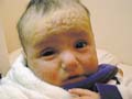
Fig. 198. Seborrheic Dermatitis
After treatment |
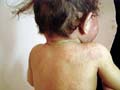
Fig. 199. Seborrheic Dermatitis |
|
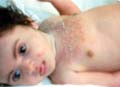
Fig. 201. Seborrheic Dermatitis |
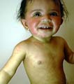
Fig. 200. Seborrheic Dermatitis
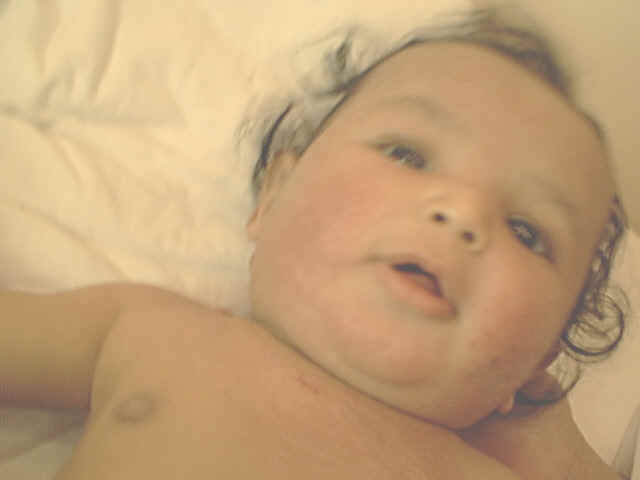
Fig.200 Seborrheic dermatitis
( Fig.200 after treatment) |
|
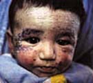
Fig. 203. Seborrheic Dermatitis |
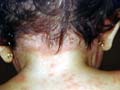
Fig. 202. Seborrheic Dermatitis |
Clinical
features
|
The sites
involved are the scalp, eyebrows, eyelids, nasolabial folds, axilla,
sternal area, umbilical, groins and the crural areas.
Skin lesions
are erythematous patches of different sizes and shapes, which are
covered by greasy scales.
Scalp
lesions present with fine greasy dandruff. The lesion may spread
beyond the hairy line of the scalp to involve the eyebrows,
nasolabial folds, face, ears and the back of scalp. In severe cases
the patches may cover the whole seborrheic areas.
|
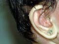
Fig. 204. Seborrheic Dermatitis
( greasy scaly lesion of the scalp&face)

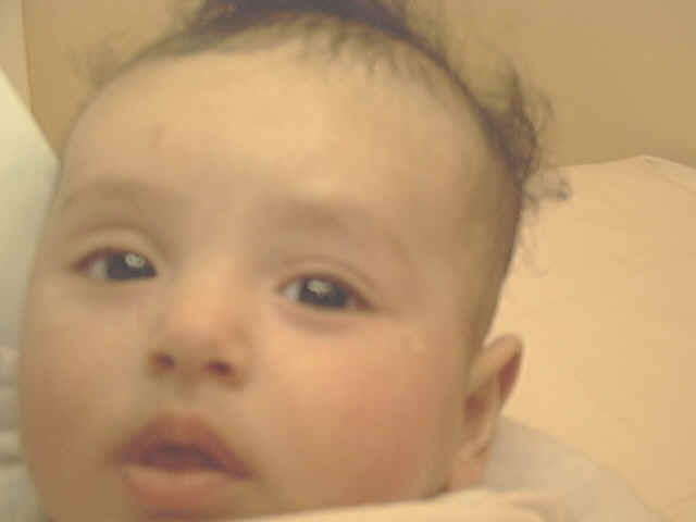
Fig.204b. Infantile seborrheic dermatitis
|
Seborrheic
dermatitis is usually accompanied by hair falling and is a common
cause of baldness in adults.
Itching is
variable but usually it is mild.
Seborrheic
dermatitis has a chronic course. The disease may become generalized
accompanied by exfoliative dermatitis or generalized erythroderma
known as "Leiner‘s disease.
Differential
diagnosis
-
Psoriasis:
psoriasis is sometimes not easily differentiated from seborrheic
dermatitis especially that of the scalp.The involvement of other
seborrheic areas and the greasy scaly patches may help in the
differential diagnosis. Psoriasis lesions do not usually extend
and exceed below the hairlines in contrast to seborrheic
dermatitis, which may extend to involve the eyebrow, eyelashes and
the face.
-
Impetigo of the
scalp and folliculitis. There is no greasy dandruff and the
lesions are usually localized.
-
Pityriasis rosea:
can be differentiated by the presence of the herald patch,
characteristic distribution of lesions around the lines of ribs ,
peripheral adherent scale and the hypopigmented patches.
-
T.
cruris and corporis:
are characterized by the active edges of the lesions, dry non
greasy scales and by detection of the causative fungi from
scrapings from the active peripheral edge of the tinea lesions.
INFANTILE
SEBORRHEIC DERMATITIS
Infantile
seborrheic dermatitis is a term used to describe a clinical
presentation, which may reflect a variety of different skin
disorders such as cradle cap, intertriginous lesions, intertrigo,
infantile psoriasis and Leiner‘s syndrome.
The
incidence of atopic dermatitis with seborrheic dermatitis is so high
that some infants with infantile seborrheic dermatitis would in any
case be expected to develop atopic dermatitis.
P. ovale is
part of the normal skin flora has a possible etiological role. This
is supported by reports of the therapeutic response of seborrheic
dermatitis to topical ketoconazole .
Treatment of
Seborrheic Dermatitis
Seborrheic
dermatitis in newborn and young children may need only mild
medications in comparison with the adult seborrheic dermatitis .
-
Skin
lesions
Mild lesions
of the skin improve usually with hydrocortisone ointment .
Moderate
cases : hydrocortisone ointment combined with tar or vioform (Lococorten
tar ,
Lococorten vioform ).
Severe cases
: Fluorinated steroids may be used taking into consideration the
side effects .
Topical
preparations in combination of antibiotics or Itraconazole can be
used in complicated cases with bacterial or fungal infections .
In most
cases of seborrheic dermatitis in infants and children , I use
non-steroid
Pufexamac in combination of antifungal and antibacterial preparation
(Flogocid , Parfenac) . This is a safe medication , effective and
gives good results .
-
Scalp
lesions of seborrheic dermatitis :
-
Mild
cases: may need simply a mild shampoo (Head & Shoulder shampoo,
Zincon shampoo) used every other day .
-
Moderate
cases : Hydrocortisone lotion ( Lococorten lotion ) , locoid scalp,
elocom lotion with a mild shampoo .
-
Severe
cases: Fluorinated corticosteroid lotions may be needed for chronic
recurrent , reluctant cases and a shampoo containing tar (Poly tar
shampoo , Zeton shampoo) .
-
Seborrheic dermatitis of the eyelids : Mild shampoo such as baby
shampoo used several times and applying hydrocortisone ophthalmic
ointment twice daily for a short period .
-
Nizoral
shampoo can be used for older children and adults for a short period
can give good results . Selenium sulfide shampoo (Selsun ) is an
effective in seborrheic dermatitis. Long use of such shampoos may
cause hair falling. Care for the eyes during washing of the scalp,
because these may cause irritation.
-
Seborrheic dermatitis of the scalp is usually complicated by
secondary bacterial infections such as folliculitis. Antibacterial
lotion such as Erythrocin (Eryderm lotion) or clindamycin (Dalacin T
lotion) can be applied twice daily to the scalp.
-
Bacterial
infections of the scalp should be treated before application of
corticosteroids lotions, where the latter are postponed till
controlling the bacterial infections. Oral antibiotics such as
cephalosporins, Erythrocin, Zithromax may be indicated in severe
extensive infections. Doxacyclines, Minocycline and Tetracyclines
are not given for children but they have good effect in adults.
CRADLE CAP
This is a
mild form of seborrheic dermatitis, affecting newborn infants. The
skin manifestations present with adherent and greasy scales
involving the vertex of the scalp , usually accompanied by milia and
comedones of the face.
Some authors
consider that type is the continuation of the vernix caseosa, which
covers the scalp of newborn after labor .
Clinical
Feature
Erythematous,
swollen and greasy yellowish adherent scales cover the scalp
especially the vertex, and eyebrows. The lesion may extend to
involve the neck , behind the ears, chest , axilla and
intertriginous areas The patches have characteristic well-defined
margins and usually symmetrical.
The areas
may become macerated, fissured and are liable to secondary bacterial
and fungal infections.
|
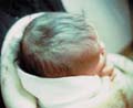
Fig. 205. Cradle cap |
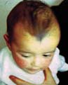
Fig. 206. Cradle cap |
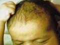
Fig. 207. Seborrheic Dermatitis( Cradle cap) |
Treatment
Management
of mild forms of cradle cap is simple by rinsing the scalp with warm
olive oil, which is left for few minuets, and the area is then
combed gently, where the scales can be easily removed leaving the
area free from scales.
The scalp
later is cleaned with mild shampoo .
Shampoos
containing selenium sulfide , salicylic acid , sulfur should be
avoided in the newborn for the possibility of toxic absorption .
For older
children: hydrocortisone alone or in combination with salicylic acid (Locosalene,
Dexalocal) can be applied gently. This may give good results.
Complicated
cases with secondary bacterial or fungal infection can be treated
accordingly using topical antibacterial preparation such as
Muperacin cream or antifungal topical preparations such as
Itraketocanazole cream .
LEINER‘S
DISEASE
Leiner‘s
disease is considered a wide spread type of infantile seborrhoeic
dermatitis with extensive erythematous and eczematous skin lesions .
Etiology
A defect of
immunity in infants with functional inadequacy of the fifth
component of complement (C5) is believed to be an important factor .
It is
believed that the syndrome of C5 deficiency and Leiner‘s disease
were one and the same .
This
syndrome may be associated with general systemic manifestations such
as diarrhea and weight loss .
Many of
these infants improved when their diet was changed to rice water and
cows‘ milk. It is also believed that breast-feeding was
responsible for the illness.
|
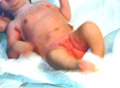
Fig.208 Leiner's disease |
Others
consider Leiner‘s disease is a purely clinical entity that is characterized by:
-
Generalized seborrheic dermatitis.
-
Intractable severe diarrhea
-
Marked
wasting and dystrophy .
-
Recurrent local and systemic infections .
-
‘Familial
Leiner‘s disease with C5 dysfunction is considered a more
dangerous variant.
Treatment
Most cases
are severe and hospitalization is necessary .
Fluid and
electrolytes balance .
Fresh plasma
.
Steroids and
antibiotics .
Dietary
supplements of vitamin B complex including biotin are important.
REFERENCES
-
Bonifazi E. Infantile seborrheic dermatitis: pathogenetic
considerations and nosological aspects. Pediatr Dermatol News
1988; 7: 16-21.
-
Yates
VM, Kerr Rei, Mackie R. Early diagnosis of infantile seborrheic
dermatitis and atopic dermatitis-clinical features. Br J Dermatol
1983; 108: 6338.
-
Prince
GE. Erythroderma desquamativa of the newborn infant. J Pediatr
1955; 47: 475-80.
-
Miller
ME, Koblenzer PJ. Leiner‘s disease and deficiency of C5. J
Pediatr 1972; 80: 879-80.
-
Evans
DIK, Holzel A, MacFarlane H. Yeast opsonization defect and
immunoglobulin deficiency in severe infantile dermatitis (Leiner‘s
disease). Arch Dis Child 1977; 52: 691-5.
Top
|