|
METHODS OF DIAGNOSING SKIN LESIONS
|
|
|
|
History taking is essentially similar to that undertaken in any general medical or surgical case. Detailed questions concerning the time of onset and duration of the skin lesion, past and family history and the medication used by the patient are important data that may help in diagnosis of skin diseases. When dealing with skin problems in infants or children, the mother gives the history or even the diagnosis sometimes. The physician should have great patience, questioning systematically and persistently. It is of prime importance to ask about all the medications that have been given to the patient whether topical or oral. Most patients consulting dermatology clinics have had their skin problem treated either by a physician, the members of the family, the pharmacist or by the advice of friends. However, when the problem persists or becomes more complicated, they seek for professional help. Past history of an allergic reaction (asthma, hay fever, urticaria, drug eruption etc.), are very important and should be recorded in the file. Detailed history concerning the type of feeding and any particular food exacerbating the skin lesions of the child should be taken into consideration. Newborn and infants are not only breast-fed but other supplements may be used as cereals, which may exacerbate their skin problem. The role of the mother is not to feed or just to fatten her baby. There are other ethical, moral, and educational duties. Supervision of her child from the anatomical, physiological and psychological point is of prime importance. The condition of the skin, whether there is dryness, or the child sweats or not, types of excreta coming out, for change of color, bloody or others all should be taken into consideration and reported to the treating physicians. These physical signs may diagnose certain systemic or genetic diseases. The physician may get valuable data from the mother, if he follows a detailed history. He can spend few minutes in questioning and directing the attention of the mother to such details and to be kept in her mind. Some of data in the history save time and effort in reaching an accurate diagnosis. The physician must not feel at all that he spent longer time with his patient. Certain medications or foodstuffs taken by the mother and pass through breast milk may have an important role in the etiology or exacerbation of dermatitis eczema reaction. The experienced physician
will not often follow a rigid line of questioning as in the type
encountered in a typical sheet form. Complete medical history of a patient
with a skin disease should trace the development of the disease as
accurately as possible; to determine those elements in the patient‘s
present or past history which may be related to the skin problem either
psychic, personal, emotional status, hereditary and environmental. In addition to the inquiry concerning familial tendencies to diabetes, cardiovascular disease and tumors, questioning concerning the familial incidences of allergic diseases is of great importance in a wide variety of skin conditions. It is not sufficient to simply ask the question "is there any allergy in the family?" Hives, eczema, hay fever and asthma should be specifically mentioned. Other common conditions in relation to skin diseases include acne, icthyosis, xeroderma pigmentosa, rosacea, baldness, psoriasis and some other congenital malformations.
As many other diseases, certain skin diseases are seen much more frequently in certain age groups. Despite the smooth and delicate appearance of a baby‘s skin, it seems to be quite similar to the adult skin. Some organs of the newborn are less well developed such as sebaceous glands, hair follicles and dermal connective tissue. There are important differences in the heat regulation between the infant and the adult skin. This is due to the different area-to-body-volume ratio, the skin vascular reactivity, and to its ability to lose heat by sweating. These very important differences should be taken into consideration especially when prescribing topical or other medications hence a child skin presents less barrier to the penetration of medications and toxins than in adults.
Environment has an important role on the pathogenesis or exacerbation of certain skin diseases in infants and young children.
Race is rarely a crucial determining factor, however this may be of considerable importance in the prognosis of certain skin diseases. Deep mycosis such as coccidomycosis may cause serious prognosis in Negroes.
Certain skin diseases such as leishmaniasis, Yaw, Pinta and Bejel are endemic in tropical areas. Contact dermatitis occurs more in areas having specific plants, trees or exposure to different chemicals. An individual who spends some time in a tropical climate, but who is not a native, is much more likely to have some very aggravated form of common skin diseases such as insect bites, fungal and bacterial infections than the native residents. Allergic rhinitis, asthma may be seen more in dusty polluted areas and in temperate zones. Some skin diseases have certain geographic distribution where the physicians of this area may be not familial with them and most of their knowledge is from literatures, while those living in the endemic area, such diseases may be faced and spot diagnosed. I have seen a young secretary lady of one of the ambassadors who was referred to me by the family physician. This lady received a heap of medications for her leg ulcers from local and other centers. The patient was under different types of topical and systemic steroids for her skin lesion (cutaneous leishmaniasis). The patient was in doubt when I told her that her problem could be solved. She was instructed to take daily injections ( 60 mg. Pentostam) for ten days. The patient used to take repeated photos for her leg ulceration. On the tenth day she visited my office cheering as there was dramatic healing of her skin lesion.
Certain diseases have seasonal peaks. Contact dermatitis and hay fever are invariably seen during the season of pollens. Many diseases including dermatitis and superficial fungal infections, miliaria become much worse during seasons of high environmental temperature, especially with high humidity climates. Papular urticaria is more common in children in summer due to insect bites. Dry skin is often worse in winter because of the low humidity of artificially heated air-conditioned houses and exposure of the skin to long hot baths. In patients with chronic recurrent skin disorders, the history of climate the time and onset of the disease, and other factors all of these are of great importance.
This is of a prime importance especially in infants and young children. The parents should be questioned about topical and internal medications given to their child. In many patients with dermatitis, it will be found that the objective changes in large part are due to the treatment that has been given to the primary skin disease. The clinical picture may be completely distorted due to some topical medications particularly with colored topical medications such as Gentian violet. This will not only mask the lesion, but it may cause maceration of the infants‘ skin especially when applied concentrated to the genital or intertriginous areas. A large number of infants and children may be seen with striae, acniform eruption, moon face, undescended testicles, wasted buttocks, thin and severe erythematous skin especially on the face due to abuse or misuse of potent steroids. Topical medications due to the base content or the medication itself or the combination with other medications such as neomycin, anesthetics, antihistamines and even steroids may cause local sensitization. Drug reactions due to antibiotics, sedatives, tonics, vitamins, laxatives, and fixed drug eruption induced by sulfonamides are not uncommon. Some beverages that contain special herbs or those in a form of herbal tea may cause severe skin reaction.
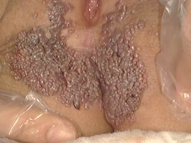
Examination should be under a good light, preferably daylight or a daylight type electrical lamp and using a magnifying lens. An assessment of a patient‘s normal skin problems can be made at a glance, with experience, but still thorough examination not only to the site of the lesion but to other parts of the body should not be neglected. The physician must get up and do something for his patient. Besides talking to him or adding humor, he should try to give confidence, courage and sympathy to the patient. He can even give "hardened pessimistic cases" an optimistic look to make his patient "cheer up", especially those who have chronic resistant dermatoses or those with psychosomatic problems. The physician should not be very serious and must not flip, but his light touch with the kindly cheery smile may dispel the gloom. Confidence of the patient or his parents towards the physician is considered of prime importance. The prescription alone sometimes will not do the whole job. The physician must impose his will on his patient, and this can be done best with graciousness, diplomacy and reason. The physician should not pay all his attention to diagnose and treat the skin lesion only. He has to know that certain skin diseases are manifestations of internal diseases. Thorough and keen examination should not neglect signs of child abuse either by the parents, or the housemaids, the drivers, or other employees working for the family. Genital areas of children should be examined for signs of local infection mainly anogenital warts, contusions, laceration of the anogenital areas. It is not uncommon that the physician may be faced with lacerated hymen, abrasions, contusions of the child‘s genitalia or even vaginal or urethral discharge or other manifestations of sexually transmitted diseases in children.
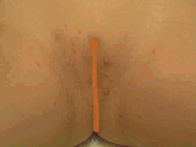
Genital contusions and mollascum contagiosum of a child
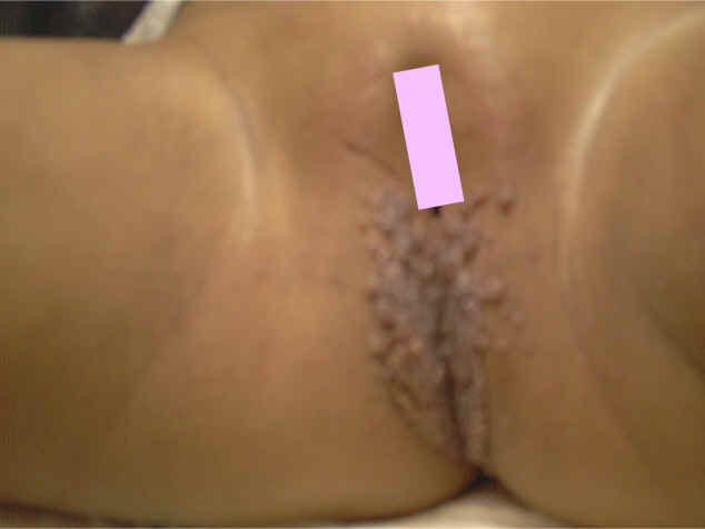
Herpes progenitalis of a child , (Possibility of sex abuse in a child) Psychological behavior and the relation of the child with his parents and other members of the family should not be neglected during examination.
PRIMARY SKIN LESIONS The primary skin lesions are the original lesions that appear as a result of different stimuli either internal or external. The different primary skin lesions seen on examination are: Macule - a circumscribed flat area of different color from the surrounding skin. Macules may become raised due to edema, where it is then called maculopapules Papule - a raised circumscribed elevation of skin. Nodule or tubercle - a solid elevation of the skin, larger than a papule. Plaque - a raised thick portion of the skin, which has well defined edges with a flat or rough surface. Erythema (redness of the skin surface) -This is the commonest primary skin lesions, which appears in most skin diseases. Erythema is due to dilatation of dermal blood vessels and edema. Blister - a skin bleb filled with clear fluid Vesicle - a small blister. Bulla - a large vesicle Pustule - a skin elevation filled with pus Cyst - a cavity filled with fluid. Nevus - hereditary skin disorders due to deficiency or excess of the normal constituents of the skin and usually defined as nevi.
Secondary skin lesions are modifications or changes of the primary lesions due to infection, trauma or due to other factors. The different secondary skin lesions are : Scales - a flake of flat horny cells loosened from the horny layer. Fine desquamation of the skin is an ordinary physiological in normal individuals due to the wear and tear. When the formation of epidermal cells are rapid or there is disturbance of normal skin keratinization pathological scaling will result. Skin scaling may be localized or generalized called as exfoliative dermatitis. Crust - dried serum seen in ruptured vesicles, pustules or bulla. Excoriation: mechanical abrasion of the skin usually caused by the fingernails in attempt to relive itching. Fissure - a crack or split in the epidermis. Erosion - an area of partial loss of epithelium of skin or mucous membrane. Ulcer - an area of total loss of epithelium of skin or mucous membrane. Atrophy - loss of thickness of the epidermis or dermis or other tissue. Lichenifecation - thickness of both prickle cell layer and horny layer with exaggeration of normal skin marks. Sclerosis - diffuse or circumscribed induration of the subcutaneous tissues. Fibrosis - the formation of excessive fibrous tissue. Abscess - a localized collection of pus in a cavity formed by disintegration or necrosis of tissue. Cellulitis - an inflammation of cellular tissue, particularly purulent inflammation of the deep dermis and subcutaneous tissue. Pyoderma - any purulent skin disease. Alopecia - localized or generalized loss of hair due to local or systemic factors. Alopecia may be primary or secondary to a local skin disease such as fungal or bacterial infections. Burrow - a small tunnel in the skin that houses a metazoal parasite, such as the scabies acarus. Comedo (nes) - a plug of keratin and sebum in a dilated pilosebaceous orifices. Hematoma - a localized tumor-like collection of blood. Ecchymoses (bruise) - a macular area of hemorrhage more than 2 cm in diameter. Petechiae - a punctate hemorrhage spots approximately 1-2 mm in diameter. Stains or pigments - local hyperpigmentation of the skin following certain skin diseases. Exfoliation - the splitting off of the epidermal keratin in scales or sheets. Hemosedroses - in stasis dermatitis. Fistula - an abnormal passage from a deep structure to the skin surface or between two structures. It is often lined with squamous epithelium. Keratoderma - a localized hyperplasia and thickening of the stratum corneum. Striae - linear lesions due to stretch of the skin, either physiological or pathological. Callus - a horny thickening of the skin. Milium - a tiny white cyst containing lamellated keratin. Vegetation - a growth of pathological tissue consisting of multiple closely set papillary masses. Papilloma - a nipple-like mass projecting from the surface of the skin. Aphtha - a small ulcer of the mucosa. The Koebner‘s or isomorphic phenomenon - is the formation of skin changes such as that of the primary skin disease by non-specific trauma as in psoriasis.
METHODS OF DIAGNOSIS IN DERMATOLOGY The skin is the exposed covering and the outer coat lining of the body surface. Skin lesions whether primary or secondary appear frankly to the naked eye. The experienced physician can immediately diagnose some skin diseases on the spot without hesitation. The patient who spent his time and money to have the opportunity to reach the physician to treat his skin problem should have appropriate and appreciated care. From the psychological point of view he may be disappointed when he finds that his problem was solved within a few minutes especially when that was done in a rush and a non-convincing way. Just a few minutes after sitting in front of his physician, the patient will be provided with a piece of paper containing the medications. He may be given the impression that the physician has finished his job and he has to leave his seat for another patient. Some patients may have doubt that his skin problem was not handled in the proper way especially those who have chronic skin diseases and have consulted different clinics. We were faced with patients saying that "these were the most expensive minutes in our life, we paid a lot of money for these few minutes". It is true that some skin diseases can be diagnosed on sight with a high degree of confidence but even in such cases, detailed history is indispensable for an effectively planned management. In spite of all of that, it is true from the physician point of view that he can diagnose most skin diseases in this way but certain skin diseases need more care and more investigations to reach a thorough and convincing diagnosis.
|
| Contents | << Previous Chapter | Next Chapter >> | Search |